If you're seeing this message, it means we're having trouble loading external resources on our website.
If you're behind a web filter, please make sure that the domains *.kastatic.org and *.kasandbox.org are unblocked.
To log in and use all the features of Khan Academy, please enable JavaScript in your browser.

AP®︎/College Biology
Course: ap®︎/college biology > unit 2, introduction to the cell.
- Organelles in eukaryotic cells
- Intro to eukaryotic cells
- Endoplasmic reticulum and Golgi bodies
- Endomembrane system
- The endomembrane system
- Mitochondria
- Mitochondria and chloroplasts
- Cell structures and their functions
Want to join the conversation?
- Upvote Button navigates to signup page
- Downvote Button navigates to signup page
- Flag Button navigates to signup page

Video transcript
Atoms Alive: A tour inside a cell
- X (formerly Twitter)
CHRISTOPHER DUNNE:
Sure, we can see organelles from a microscope, but that doesn't tell us what they do. To do that, we need to get a little brutal. First, we grab some tissue, some ordinary spinach and add it to a household blender. Just give that a quick buzz. Now, this has created a cell soup, which we'll now take to the centrifuge. First, we add our cell soup in a tube. This spins very, very fast and works like supergravity. Inside, the heavy particles fall to the bottom in a pellet. To get it going, we add a safety lid… ..close the lid and press the go button. Now that the centrifuge has finished, we can remove our tube. Now you can see here, a pellet has been formed by the heaviest particles falling to the bottom of a tube. We can conduct chemical tests on this, to find out how they react. That's how we know the chloroplast are involved in photosynthesis because we can separate them. That's how we know that mitochondria's involved in respiration 'cause we can separate them. And that's how we know that DNA is mostly found in a nucleus and so on. In fact, we can use an ultra-fast centrifuge to even separate molecules of different sizes.
So what do we know about the complex and active interior of a eukaryotic cell? Let's take an animated tour. First, we approach the plasma membrane, which, as we pass through, we see as a lipid bilayer. Lots of special proteins stick through it, acting as selective receptors and pathways to move molecules in and out. Small molecules like water sneak through everywhere. It's very flexible and self-sealing, so it can engulf particles and carry them inside. Inside, it's a mass of strands and organelles, which we've coloured to make them easier to distinguish. In reality, they're nearly all transparent and we're not moving through empty space, but through a thick and soupy cytoplasm. It's laced through with fibres of protein. The thin green ones are called microfilaments and the thicker blue ones are microtubules because they're hollow. Both kinds make up a kind of skeleton for the cell called a cytoskeleton. But this skeleton is incredible. It's a shape-changer. When you see a cell contract or bend, you're looking at the effect of protein molecules in the cytoskeleton, using energy to change shape or to slide across each other. Tiny molecular motors anchored in the cytoskeleton power beating cilia or flagella. In fact, muscle power on whatever scale is the cytoskeleton at work. Back in our cyber cell, we run into more strange objects. Inside the mitochondrion is where the oxygen-consuming reaction that produces most of the cell's energy takes place. The folded bag of the Golgi apparatus is like a post office for special proteins. It packages them and sends them to specific places. For example, remember the food particle we saw come in? To digest its contents, a package of digestic enzymes is sent to fuse with it. Hundreds of different protein packages have jobs to do in the cell. The basic proteins are made here in the endoplasmic reticulum. These little dark dots called ribosomes are the sites of protein synthesis. Some are also found floating free in the cytoplasm. Then, hidden behind the layers of endoplasmic reticulum, is the nucleus, protected by its own nuclear membrane. Inside here, we see lots of fibrous threads and a nucleolus, where ribosomes are being made. It's the threads called chromatin which holds the crucial molecules of the cell's existence. The chromatin proteins are like spindles and wrapping paper for the nano thin but immensely long molecules of DNA, from which all the instructions for cell chemistry are drawn. A human cell has as much as 2m of DNA in every nucleus. When the cell starts to divide, these chromatin proteins draw the DNA even tighter into the characteristic shapes of chromosomes. Of course, once biochemists realised that DNA ultimately controlled all the cell processes, including this feat of cellular reproduction, they set to work to find out how.
SUBJECTS: Science
YEARS: 9–10
Take an animated tour inside a Eukaryotic cell.
Discover the different compartments (organelles) and structures.
Travel across the plasma membrane and see how different particles enter a cell.
Learn about the cytoskeleton, which is the network of fibres that help maintain the shape of the cell.
See how the DNA is packaged inside a central compartment called the nucleus.
Things to think about
- 1. Imagine the inside of a cell. Do you think there is empty space in between the cell's structures? Or do you think it consists of something else? If the DNA inside a single human cell was stretched out, how long would it be? How do you think all this DNA is packed inside a cell?
- 2. As you travel across the plasma membrane of the cell, can you see water molecules readily traversing the membrane? Describe how larger particles move into a cell. Organelles are suspended in the cytoplasm, the fluid that fills a cell. See the jungle of strands called the cytoskeleton. It is made up of what two kinds of protein fibre? Inside the nucleus is a mass of threads called chromatin. What is chromatin?
- 3. List the different organelles inside a cell and research their functions. It might be helpful to view animations of cells. View websites that show the structures of cells such as 'Cells Alive'.
- 4. To explain the function of organelles in a Eukaryotic cell they are sometimes compared to a factory. Research the function of different organelles and consider how their roles might resemble different departments within a factory. For example, the nucleus might be like the chief executive officer who controls everything.
Date of broadcast: 13 Aug 2002
Metadata © Australian Broadcasting Corporation and Education Services Australia Ltd 2012 (except where otherwise indicated). Digital content © Australian Broadcasting Corporation (except where otherwise indicated). Video © Australian Broadcasting Corporation (except where otherwise indicated). All images copyright their respective owners. Text © Australian Broadcasting Corporation and Education Services Australia is licensed under a Creative Commons Attribution-ShareAlike 4.0 International License (CC BY-SA 4.0).
Atoms Alive: Introduction to cells
Atoms Alive: Cells and energy
Atoms Alive: Lipids form cell membranes
- News/Events
- Arts and Sciences
- Design and the Arts
- Engineering
- Global Futures
- Health Solutions
- Nursing and Health Innovation
- Public Service and Community Solutions
- University College
- Thunderbird School of Global Management
- Polytechnic
- Downtown Phoenix
- Online and Extended
- Lake Havasu
- Research Park
- Washington D.C.
- Biology Bits
- Bird Finder
- Coloring Pages
- Experiments and Activities
- Games and Simulations
- Quizzes in Other Languages
- Virtual Reality (VR)
- World of Biology
- Meet Our Biologists
- Listen and Watch
- PLOSable Biology
- All About Autism
- Xs and Ys: How Our Sex Is Decided
- When Blood Types Shouldn’t Mix: Rh and Pregnancy
- What Is the Menstrual Cycle?
- Understanding Intersex
- The Mysterious Case of the Missing Periods
- Summarizing Sex Traits
- Shedding Light on Endometriosis
- Periods: What Should You Expect?
- Menstruation Matters
- Investigating In Vitro Fertilization
- Introducing the IUD
- How Fast Do Embryos Grow?
- Helpful Sex Hormones
- Getting to Know the Germ Layers
- Gender versus Biological Sex: What’s the Difference?
- Gender Identities and Expression
- Focusing on Female Infertility
- Fetal Alcohol Syndrome and Pregnancy
- Ectopic Pregnancy: An Unexpected Path
- Creating Chimeras
- Confronting Human Chimerism
- Cells, Frozen in Time
- EvMed Edits
- Stories in Other Languages
- Virtual Reality
- Zoom Gallery
- Ugly Bug Galleries
- Ask a Question
- Top Questions
- Question Guidelines
- Permissions
- Information Collected
- Author and Artist Notes
- Share Ask A Biologist
- Articles & News
- Our Volunteers
- Teacher Toolbox

show/hide words to know
ATP: adenosine triphosphate. ATP is the energy-carrying molecule of all cells...... more
Cellulose: the structural material found in the cell wall in most plants. Cellulose is used to make many products, including paper and cloth... more
Chromosome: a long, thread-like molecule made of the chemical called DNA (deoxyribonucleic acid) that is held together with special proteins and is visible (with strong microscopes) during cell division... more
Molecule: a chemical structure that has two or more atoms held together by a chemical bond. Water is a molecule of two hydrogen atoms and one oxygen atom (H2O)... more
Organelle: " little organ ". An internal organ of a cell... more
Phospholipid: is a special kind of lipid that is made up of two fatty acid chains. These phospholipids are present in the plasma membrane of any cell... more
Photosynthesis: a set of chain reactions that convert light energy into chemical energy. Photosynthesis also produces energy-rich carbohydrates like starch. Photosynthesis occurs in the chloroplast of a plant cell... more
Tomography: process used to make a tomogram (picture) which is a two-dimensional slice of a three-dimensional object. A computer can then be used to build a three-dimensional image of the object by stacking the tomograms together.
Do All Cells Look the Same?
Cells come in many shapes and sizes. Some cells are covered by a cell wall, other are not, some have slimy coats or elongated structures that push and pull them through their environment. Some cells have a thick layer surrounding their cell. This layer is called the capsule and is found in bacteria cells.
In our body there are many different kinds of cells. We are made up of about 200 different types of cells. Our body also has non-living materials such as hair, finger nails, and the hard part of teeth (enamel). All these materials are made up of dead cells or other minerals.
What Are the Parts of the Cell?
Have you ever wondered what the inside of a cell looks like? If you think about the rooms in our homes, the inside of any animal or plant cell has many similar room-like structures called organelles. Each organelle is a place where specific jobs are done.
Plant and animal cells have many of the same organelles. But in some cases, the organelles in cells are different. For example, in plant cells, there are more types of organelles than are found in animal cells. And fungal cells have organelles not found in any other cell type.
Below are some names and descriptions of organelles commonly found in certain cells. There is also an interactive cell viewer and game that can be used to learn about the parts of animal, plant, fungal, and bacterial cells. Archaea cells are very similar to bacterial cells, so have not been included separately. An introduction video is now available to see how the game is played.

Read more about: Building Blocks of Life
View citation, bibliographic details:.
- Article: Parts of the Cell
- Author(s): Shyamala Iyer
- Publisher: Arizona State University School of Life Sciences Ask A Biologist
- Site name: ASU - Ask A Biologist
- Date published: September 27, 2009
- Date accessed: May 28, 2024
- Link: https://askabiologist.asu.edu/cell-parts
Shyamala Iyer. (2009, September 27). Parts of the Cell. ASU - Ask A Biologist. Retrieved May 28, 2024 from https://askabiologist.asu.edu/cell-parts
Chicago Manual of Style
Shyamala Iyer. "Parts of the Cell". ASU - Ask A Biologist. 27 September, 2009. https://askabiologist.asu.edu/cell-parts
MLA 2017 Style
Shyamala Iyer. "Parts of the Cell". ASU - Ask A Biologist. 27 Sep 2009. ASU - Ask A Biologist, Web. 28 May 2024. https://askabiologist.asu.edu/cell-parts

Building Blocks of Life
Coloring Pages and Worksheets
Animal Cell
Bacterial Cell
Fungal Cell

Be Part of Ask A Biologist
By volunteering, or simply sending us feedback on the site. Scientists, teachers, writers, illustrators, and translators are all important to the program. If you are interested in helping with the website we have a Volunteers page to get the process started.
Share to Google Classroom
- Workshops & Institutes
- Curriculum Index
- Research Opportunities
Sign In with Google
Create an Account
Stay informed! Sign up for our newsletter. We will never send you spam or sell your information.
Please verify that you are a teacher
Sign up with Google
Why should I sign up?
Even without an account, you’ll still have free access to most of the award-winning content on Teach.Genetics. Creating an account will give you access to additional content and tools.
Reset Password Email: Reset Password Email
- Amazing Cells
Here you’ll find a number of multimedia and paper-based classroom resources, featuring dynamic and realistic depictions to help you explore the inner-most workings of cells.
The Cells In Context section describes a sequence of related resources that work together as a middle school cell biology unit.
The Cells Communicate section describes additional resources designed for high school.
Interactive Tools
This magical microscope lets viewers jump between levels of magnification from organ systems to cells.
This dynamic tour features 3 different cell types, each with animated depictions of organelles working together to carry out basic life functions. Explore the functions to learn the name of each cell structure and its role in the cell.
Use an interactive slider to compare the relative sizes of objects, cells, organelles, molecules, and other biological structures.
Image Files
We offer most of the graphics from the print-based materials below as image files. You can download activity-specific bundles of images as ZIP files and use them with your favorite tools. Plug them into digital whiteboards (like Jamboard), slides, documents—anywhere you can drop a jpeg file!
Cells in Context Image Files
CELLS IN CONTEXT - Suggested Lesson Sequence
This middle school unit’s resources are designed to be used in any order, with or without outside lessons. However, we hope you will consider the suggested sequence below. It pulls together the unit’s resources in a way that illuminates the connection between structure and function and examine how cells work together in systems.
The unit’s materials offer an in-depth exploration of specialized cell types. Student pairs can follow one cell type through several activities, or they can learn about multiple cell types. Three cell types (airway, intestine, and leaf) appear in all the key modeling activities: Mystery Cell Model, Teaming with Cells, Hijacked Cells!, and Hijacked Teams! Mystery Cell Model features two additional cell types: neuron and plant root cell.
- Three-dimensional
- US Middle School level (ages 12-14)
- Flexible: use in sequence as a complete unit or integrate with other curriculum materials
- Consistent visual language: structures are depicted similarly throughout
- Uses models to visualize cell structure and function
NGSS Connections
NGSS Phenomena
Is it Alive?
How do you know if something is living or not? Students look at objects on illustrated cards (24 total) and determine whether they are living or non-living. Several tricky examples are included (such as seeds and wood) to encourage discussion about what exactly it is that makes something alive. Regardless of the criteria your students use, you’ll want to underscore that all living things are made of cells.
Have students sort cards independently, then lead a class discussion.
- There are criteria that some use to determine if something is living or not, but some examples are tricky.
- Living things are made of cells.
- Cells are the smallest unit that can be said to be alive.
Printable Object Cards with Teacher Guide (pdf) — Make one set per pair or small group (card sets can be re-used)
Is It Alive? (online version)
Mystery Cell Model
How do cells carry out the basic functions of life? Students label the structure & function of organelles on a cell model—with a slight twist. There are 5 models to distribute, each depicting a specialized cell with some parts that are unique to its function. While labeling the functions of their cell organelles, students compare their cells to find organelles that are: (1) common to all cells, and (2) unique to each cell type. Finally, they deduce their cell’s identity.
Note: The airway, intestine, and leaf cells appear in other modeling activities: Mystery Cell Model, Teaming with Cells, Hijacked Cells!, and Hijacked Teams! You may choose to work with any number of cell types, as appropriate for you students. You may wish to have each student follow the same cell type throughout, as we have found this to be a little quicker.
Have students work individually or in pairs. See Teacher Guide for details.
- Before — to introduce organelles and their function.
- During — as a whole-group or individual reference to help students identify and label common organelles.
- After — as a check to make sure students labeled their organelles correctly.
- Within cells, special structures carry out particular functions.
- All cells have many of the same basic structures, yet they also have differences that allow the cells to perform specialized roles.
Prep time: 30 minutes
Class time: 45 minutes
- Computers with internet and headphones
- Generic cell models and copies of the Most Cells Have These Parts sheet
Teacher Guide (pdf)
Cell Models:
- 8.5 x 11 (pdf) or 11 x 17 (pdf) —
Structure-Function Organizer (fillable pdf)
Most Cells Have These Parts (pdf)
Inside a Cell (interactive)
Coffee to Carbon
How big are cells? Put the relationship between cells, organelles and molecules in to perspective. Using copy-and-cut cards, students place biological structures in order by their relative size from largest to smallest.
Distribute shuffled sets of object cards to student groups and instruct them to arrange the objects pictured in order from largest to smallest. Ask students to compare the order of their cards with another group and discuss any discrepancies. Use Cell Size and Scale to check answers.
- Understand the relative size of microscopic biological structures
Prep time: 10 minutes
Class time: 20 minutes
Cell Size and Scale (interactive)
(Optional) Real Cell Gallery
Cells your textbook never dreamed of
In biology, there is always an exception to the rule. Real and illustrated examples of some interesting prokaryotic and eukaryotic cells underscore the specialized functions of cells as well the things all cells have in common.
20 - 30 minutes
Computers with internet access
Real Cell Gallery (interactive)
Introduce Levels of Organization
Using this online interactive as a demo, zoom in and orient students to cells and their context in higher levels of organization.
Navigate to the Virtual Microscope. Project and orient students to the levels of organization that they will be using throughout the unit: organ system, organ, tissue, and cell.
- Living things are made of many different numbers and types of cells.
5 - 10 minutes
Teaming with Cells
How do groups of cells work together to carry out functions in organisms? Students examine labeled illustrations and construct explanations for how a particular cell type—and the tissue and organ that it is part of—works with others to help an organism function.
Builds on Mystery Cell Model — Model a cell, then learn how it works with other cell types in a tissue and beyond!
Have students use either printed illustrations or the virtual microscope to explore four levels of organization (cell, tissue, organ, organ system).
If students have trouble finding the words for their organizers, you could either point them to the yellow boxes on the cards or provide a word bank.
- Cells form tissues and tissues form organs specialized for particular body functions.
Class time: 45 - 60 minutes
Student Organizer (fillable pdf)
Printable Illustrations (pdf) — Make one set per pair or small group (card sets can be re-used)
Online alternative for viewing illustrations: Virtual Microscope (interactive)
Hijacked Cells!
This and the next activity explore what happens when an organism’s cells are disrupted by pathogens, using the following pathogen/cell type pairs:
- Influenza virus & Airway cell
- E. coli bacteria & Intestine cell
- Tomato spotted wilt virus (TSWV) & Leaf cell
You may choose to work with any number of the infections. If you plan to work with more than one, we have found that it is a little quicker to have students follow the same cell type all the way through.
Hijacked Cells! builds on Mystery Cell Models . Students model the process a specific pathogen uses to infect a cell. They identify which organelles the pathogen uses and how it disrupts the cell’s function.
Have students work individually, in pairs, or in small groups.
- Distribute matching sets of Mystery Cell Models (linked above), Pathogen Cut-Outs, and Modeling Instructions.
- Have students follow the instructions, taping the cut-outs onto the cell models only where the instructions say to (some parts should not be taped down).
- Have students work through the Organizer, using the instructions there.
- Pathogens interrupt the normal function of particular cells.
Prep time: 15 minutes Class time: 45 minutes
Copies, tape, scissors (if students need to prepare their own cut-outs)
- How are the 3 essential functions of a cell (instructions, energy, container) affected in the case of each of the pathogens?
- In the case of E. coli , one of the essential functions is not affected. Which one, and what does the pathogen do instead to spread the infection?
- In the case of a virus, one of the essential functions is not affected. Which one and why?
- Are viruses and bacteria living? Why or why not?
- What are the top reasons the pathogen you modeled needs a host cell?
Use the optional Structure & Harm cards to help students find the right words. Provide one or both sets of cards for students to tape onto their organizers.
If you’re using the fillable pdf, you could use the information from the cards to make a word bank.
Act out a class-wide infection of either Influenza or TSWV. One student/group starts the infection. Their cell becomes the factory. After going through the virus infection cycle, they give their mature virus particles to another group. Now 2 groups are making virus and infecting others. Continue until all groups are infected.
Printable Pathogen Cut-Outs (pdf) —
Printable Modeling Instructions (pdf) —
Student Organizer (fillable pdf) — Make one per student
Structure & Harm Cards (pdf) — optional
Hijacked Teams!
Students follow their infections to the next level. Building on what they learned in Teaming with Cells, students see how pathogens disrupt tissues, organs, and systems. They piece it together to understand how exactly pathogens make you sick.
- Distribute sets of infection cards and organizers. Have students follow the instructions to complete the organizer. It may be useful for students to refer to the healthy structures and functions shown in the Teaming with Cells illustrations or Virtual Microscope
Note: Not all symptoms can be traced back to the cell level, but at least one can for each pathogen/cell type pair (see answer key); students will need to grapple with the information in the infection cards to identify which one it is.
- When cell function is disrupted, tissue function is disrupted.
- The symptoms of a disease or illness are a direct result of disrupted cell, tissue, and organ function.
Copies Student organizers from earlier activities may be useful
- Often it is the immune system that kills an infected cell. Why would it be bad for a pathogen to kill the cell? Why would an organism need to kill its own cells?
- What strategy or strategies does the pathogen use to spread to other individuals?
Have students go back through the Hijacked Teams! cards and look for instances where multiple symptoms can be traced back to one effect at the cellular or tissue level.
Play Pathogen Attacks , a board game where teams of cell specialists apply their new knowledge of pathogens and symptoms.
Hijacked Teams! Cards (pdf) Includes card sets for 3 infections. (can be re-used)
CELLS COMMUNICATE
Designed for high school students, the materials in this section build on the middle-school-level materials above. The lessons here explore cell communication from a molecular perspective.
Build-A-Membrane - Advanced
Cut, fold, and tape biomolecules to create a three-dimensional cell membrane with embedded proteins.
Have students (individually or in pairs) build membrane segments, then put them all together to form a large membrane.
- Membranes have proteins embedded in them.
- Membrane-embedded proteins allow cellular signals and other molecules to pass through the membrane.
Class time: 30 minutes
Copies, scissors, tape
- A cell is enclosed and defined by an outer membrane.
- Integral proteins, which extend through one of both layers of the phospholipid bilayer
- Proteins attached to lipid molecules that anchor them to the membrane
- Receptor proteins, which transmit signals across a membrane
- Transporter and channel proteins, which form pores through the membrane that can open and close to let specific molecules through
- Membranes also organize the interior of a cell. They wrap around compartments / organelles
- Phospholipids spontaneously arrange themselves into membranes
Student Instructions and Cut-Outs (pdf) Make one per student or pair
The Fight or Flight Response - Advanced
Watch how cell communication carried out by molecular signals bring about physiological change during the fight or flight response.
Project to the class or have students explore individually in pairs.
- Cell communication is a multi-step process.
- Cells communicate via signaling pathways made of interacting components.
- Components of cell signaling pathways sometimes change shape as a result of their interaction (conformational change)
15 - 30 minutes
Projector and speakers or individual student computers
The Fight or Flight Response (video)
(optional) Play-By-Play (pdf) - A scene-by-scene guide to the molecular interactions taking place in the video.
Related Resource: How Cells Communicated During Fight or Flight (web page) - An in-depth look at one axis of cell communication during the fight or flight response.
Pathways with Friends - Advanced
Directed by instructional cards, students kinesthetically model cell communication by acting as components in a cell signaling pathway.
- Create a space in which students can move freely.
- Each person will be given a card.
- Do not let others what know what your card says.
- When prompted, follow the instructions on the card to create a cell signaling pathway.
- Distribute one set of Cell Communication Cards to each group, and ask the students to choose a card from their set.
- Once every student has a card, prompt the groups to begin by following instruction #1 on their card.
- Next, instruct your students to follow instruction #2 on their card.
- When each group is finished, project to the class the Cell Signaling Steps diagram, summarizing the steps the students just demonstrated. Discuss the activity and how it models signaling pathways in the cell.
Copies, projector
- What happened?
- How did you recognize where to go?
- How does this model cell communication?
- What effect did joining the pathway have on you? (Looking for something to indicate conformational change.)
- What problems did you encounter?
- What would have happened if someone did not do their job (follow instructions) or were not there?
Instructional Cards (pdf) - includes Cell Signaling Steps diagram
Dropping Signals - Advanced
Students drag and drop to see how various signals affect a selection of cell types.
Have students work individually or in pairs to explore the interactive. Students can record information on an optional student organizer.
- There are different types of cells, and different types of signals.
- Cells respond differently to signals depending on cell and signal type.
Student computers with internet access (optional) copies
Student Sheet (fillable pdf)
Dropping Signals
Troubleshooting
About These Resources
This work was supported by Science Education Partnership Awards (Nos. R25RR023288 and 1R25GM021903) from the National Institute of General Medical Sciences of the National Institutes of Health.
The contents provided here are solely the responsibility of the authors and do not necessarily represent the official views of the National Institutes of Health.
- Molecular Nature
- Natural history
- Short Observations
- Human Nature
- Global Patterns
- Cosmological
- NatureDocumentaries.org
Share this post
Posted by Uzay Sezen on March 9, 2019 at 8:12 pm
A Virtual Tour of a Cell – XVIVO Scientific Animation (2018)
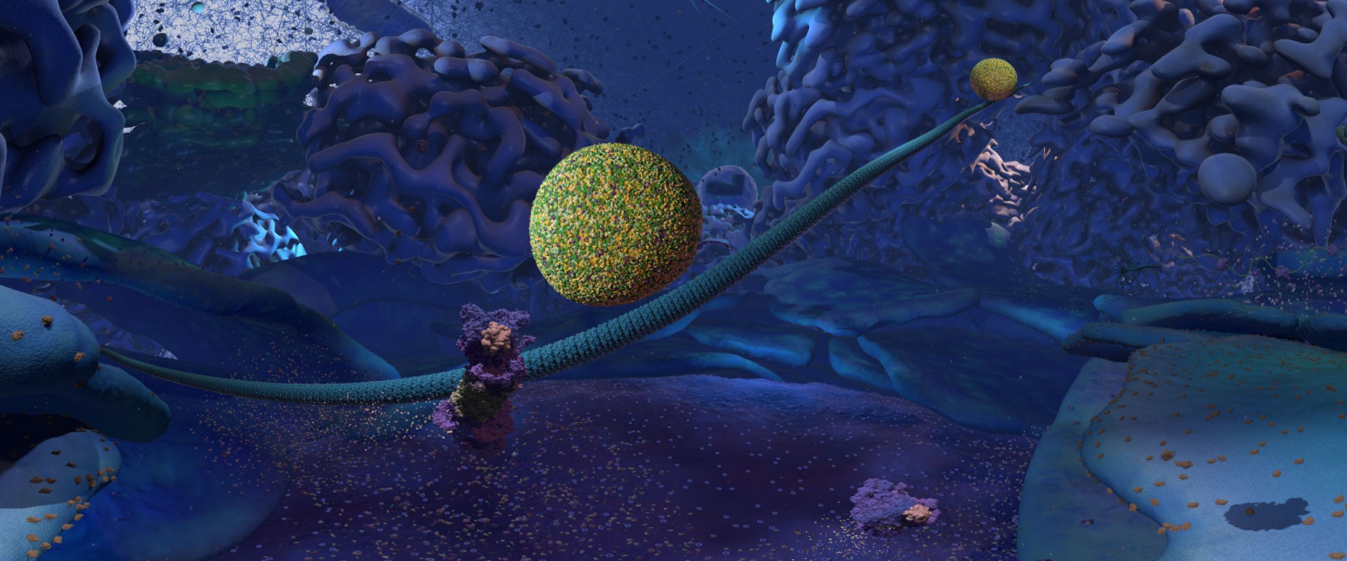
The Cellscape is one of the many fascinating scientific visualizations created by XVIVO Scientific Animation Studio. The project was supported by Google Making and Science Team . The creators did a wonderful job in introducing us to the major parts of the cell visible to a viewer in the cytoplasm in a virtual environment.
Conjuring the physico-chemical structures such as proteins and enzymes inside a cell is a rather difficult thing to do. The textbooks and diagrams, don’t do a justice since they inevitably oversimplify the ultra-dynamic, complex and mesmerizingly interesting nature of the cell. The Cellscape takes a field trip inside the cell designed to be explored with a Google Cardboard VR headset . The immersive experience helps learners to perceive cells with internal components and understand how they all function collectively.
Make your own VR viewer
As scientists determine high resolution molecular structures of subcellular compartments visualizations are increasingly becoming sophisticated . This visualization is rather animal-centric but you can check out the photosynthetic chromatophores of purple bacteria if you want to get a taste for an autotrophic cell.
The structures highlighted in Cellscape include microtubule highways, kinesin motor proteins traveling on them carrying membrane vesicles and the membrane bound organelles characteristic of eukaryotic organisms such as the endoplasmic reticulum, the Golgi apparatus and mitochondria. We also see some large macromolecules that work as molecular machines such as the ribosome -a key player in the Central Dogma of biology- and the proteasome -shredder of proteins tagged for degradation-.
Proteins are transported from the endoplasmic reticulum to the Golgi apparatus by vesicles traveling along the microtubules. Protein glycosylation, initiated in the endoplasmic reticulum, is completed inside the Golgi apparatus. Fully glycosylated proteins are transported from the Golgi apparatus to the plasma membrane. When a vesicle fuses with the plasma membrane, proteins inside the vesicles are secreted, and proteins embedded in the vesicles membrane diffuse in the cell membrane. Protein glycosylation is a very dynamic and complex process which modifies and makes proteins ready for diverse functions. For instance, the antifreeze glycoproteins in the Antarctic icefish and arctic cod fishes enable survival and adaptation in such harsh polar environments. Incredibly both species have lost their hemoglobin genes.
The following illustration summarizes the traffic between endoplasmic reticulum and golgi including some of the important glycosylation steps within the membrane folds:
image source
Leave a Comment Click here to cancel reply.
This site uses Akismet to reduce spam. Learn how your comment data is processed .
Recent Posts
- Antifreeze Proteins in Virtual Reality – Nanome (2022)
- A Yellow-throated Warbler Collecting Nesting Material
- Cornell University Hawk Camera
- Albatross Nest Live from New Zealand
- Decorah Eagle Cam – Nesting Season 2024
Recent Comments
- Este es el aspecto que tenía el bosque más antiguo conocido, hace 390 millones de años - LUDD on Devonian Fossil Forest of Gilboa
- Chase Tag Championship and the Evolutionary Dynamics of Predator Prey Interaction | Natural History Nature Documentary on Hungry Polar Bear Ambushes Seal | The Hunt | BBC Earth (2017)
- Field trip to SW Minnesota – Geological Society of Minnesota on Geological History of the Continents/Paleomap Project – Christopher Scotese (2015)
- Can Money Stop Deforestation? – The Economist (2023) | Natural History Nature Documentary on 7 Ways Blockchain Can Stop Climate Change & Save The Environment – WEF (2017)
- Can Money Stop Deforestation? – The Economist (2023) | Natural History Nature Documentary on A Satellite-eye View of Three Decades of Deforestation in the Amazon
- January 2024
- November 2023
- September 2023
- February 2023
- October 2022
- September 2022
- August 2022
- February 2022
- December 2021
- September 2021
- August 2021
- February 2021
- December 2020
- October 2020
- August 2020
- February 2020
- January 2020
- December 2019
- October 2019
- September 2019
- August 2019
- February 2019
- January 2019
- December 2018
- November 2018
- October 2018
- August 2018
- February 2018
- January 2018
- December 2017
- November 2017
- October 2017
- September 2017
- August 2017
- February 2017
- January 2017
- December 2016
- October 2016
- September 2016
- August 2016
- February 2016
- January 2016
- December 2015
- November 2015
- October 2015
- September 2015
- August 2015
- February 2015
- January 2015
- December 2014
- November 2014
- October 2014
- September 2014
- February 2014
- January 2014
- December 2013
- November 2013
- October 2013
- September 2013
- August 2013
- January 2013
- December 2012
- October 2012
- September 2012
- August 2012
- February 2012
- January 2012
- BBC documentaries
- Ecological Documentaries
- Nova Documentaries
- Technical Notes
- Wildlife Documentaries
Copyright © 2024 Nature Documentaries. All Rights Reserved.
- The Open University
- Guest user / Sign out
- Study with The Open University
My OpenLearn Profile
Personalise your OpenLearn profile, save your favourite content and get recognition for your learning
About this free course
Become an ou student, download this course, share this free course.

Start this free course now. Just create an account and sign in. Enrol and complete the course for a free statement of participation or digital badge if available.
1 Introducing the cell
Studies into the function and appearance of cells reveal the presence of great diversity, which is particularly striking among eukaryotic cells. Even very different types of eukaryotic cells, however, exhibit many common structures and functions. This mix of uniformity and diversity is also reflected in the organisation within cells. This course looks in more detail at the interior of cells - their subcellular structure, including their 'ultrastructural' features (sometimes called 'fine structure'), which are visible using electron microscopy. Knowledge of the subcellular components of cells and how these components are arranged is fundamental to your understanding of how cells perform their functions: that is, how cells 'work'.
Schematic diagrams of typical animal and plant cells are shown in Figure 1a and b. You do not need to study this figure in detail now; it will be referred to again during the course of this course.

The diagrams are three-dimensional representations of the cell's internal structure. Near the centre in both cells is the nucleus, which contains a small nucleolus. The nucleus is surrounded by a double membrane, or envelope, with pores in its surface. Outside of and continuous with the nuclear envelope are the membranous sacs that make up the endoplasmic reticulum. The endoplasmic reticulum nearest the nucleus is studded with ribosomes and is described as rough; while further out from the nucleus is the smooth endoplasmic reticulum, which lacks associated ribosomes. Other membranous sacs present in both cells make up the Golgi apparatus. Also present in the cytosol are numerous free ribosomes, small spherical structures called peroxisomes and several mitochondria; the mitochondria have an outer membrane and also a highly convoluted inner membrane. Both the animal cell and the plant cell also contain cytoskeletal components - microfilaments, intermediate filaments and microtubules; and both cells are bounded by the cell membrane. Present in the animal cell' but not shown in the plant cell, are lysosomes and also a pair of small cylindrical organelles collectively called the centrosome. The plant cell differs from the animal cell in having a cell wall outside the cell membrane, which is punctuated by connections called plasmodesmata - these are channels, lined by cell membrane, that pass through the cell walls of adjacent cells. Finally, the plant cell contains several chloroplasts, oval organelles containing stacks of green, chlorophyll-containing flattened sacs called grana; the grana are linked by membranes called grana lamellae.
Before beginning your study of cell components, some of the main techniques used to study the interior organisation of cells are briefly outlined.
INTERACTIVE
Cell explorers.
The evil Mr. X released a dangerous virus and hid the antivirus formula inside the organelles of a cell! Special agent Luca has volunteered to be micronized and enter the cell. Help her search for the formula while Hooke, her partner, provides information about cell structures and their function!
Pre-Discussion Questions:
- What is a cell?
- What is an organelle?
- What are the major parts of the cell?
- What is a virus (if you don't know the definition, an example will do)?
- What functions does a cell need to perform to survive?
Post Discussion Questions:
- According to the game, what are five life functions of cells?
- What is the difference between a prokaryotic and eukaryotic cell, and why did she think that this one is probably eukaryotic?
- Explain what the following cell structures do: cell membrane, Golgi apparatus, smooth endoplasmic reticulum, rough endoplasmic reticulum, ribosome, vacuole, lysosome, cytoplasm, mitochondria, and nucleus?
- Why does the cell create many mitochondria?
- Have a debate - which organelle visited in the game do you think is the most important?
- Use the images in the game to sketch the shape of each organelle?
Media Credits
The audio, illustrations, photos, and videos are credited beneath the media asset, except for promotional images, which generally link to another page that contains the media credit. The Rights Holder for media is the person or group credited.
Partner Organization
Last updated.
October 19, 2023
User Permissions
For information on user permissions, please read our Terms of Service. If you have questions about how to cite anything on our website in your project or classroom presentation, please contact your teacher. They will best know the preferred format. When you reach out to them, you will need the page title, URL, and the date you accessed the resource.
If a media asset is downloadable, a download button appears in the corner of the media viewer. If no button appears, you cannot download or save the media.
Text on this page is printable and can be used according to our Terms of Service .
Interactives
Any interactives on this page can only be played while you are visiting our website. You cannot download interactives.
Related Resources
A Tour of the Cell: Crash Course Biology #23
< Back to Crash Course Series
The cell is the basic unit of life, and our understanding of it has advanced as science, and the tools available to scientists, has advanced. In this episode of Crash Course Biology, we’ll take a look at the difference between prokaryotic and eukaryotic cells, take a guided tour of the eukaryotic cell, and learn why most cells are small. We’ll explore the eukaryotic cell’s surprising beginnings through an endosymbiosis that occurred about 1.5 billion years ago.
Spanish Video
Related BioInteractive Resources
- Cystic Fibrosis Mechanism and Treatment
- Root Movement
VIRTUAL REALITY CELLSCAPE EXPERIENCE
As early as the 3rd century BC, scientists dedicated their lives to understand our innermost workings. But, thanks to virtual reality, you don’t need to be a scientist to discover how the body works on the cellular level. XVIVO Scientific Animation and YouTube educator, Tyler DeWitt coupled with a generous grant from Google Making & Science Team have created Cellscape: an immersive, educational 3D virtual reality tour that takes a look inside a human cell.
What is Cellscape?
Virtual Reality (VR) promises to be a revolutionary device in education, as it allows individuals to “experience” processes and complex systems that until now could only be explained in video or textbooks. For the immersive 3D experience, view Cellscape in a VR headset, such as Google Cardboard. Alternatively, Google Chrome and the YouTube smartphone app allow users to navigate the environment.
You’re made of about 40 trillion cells. Dive into one of them, and explore the amazingly beautiful, complex, and dynamic microscopic world!
Tyler DeWitt on Cellscape:
“I’ve taught Biology for many years, and no matter whether I’m teaching elementary school or college, students have tremendous difficulty picturing the physical world inside of a cell. The cell is an incredibly dynamic place, and teachers try explain all the things that are going on: they have textbooks and diagrams, but it’s sort of like trying to describe New York City to students when all you’ve got are some crude hand-drawn maps. I always felt that if I could just take students on a field trip inside the cell, they could look around and see things, and suddenly, it would all make sense. Cellscape is an attempt to do just that. Put on a VR headset, and you’re suddenly inside this beautiful amazing alien landscape that is literally right inside you. Finally, students can really picture cells, know the components inside, and understand how they all function together. And most importantly, they can start to ask the right questions.
Cellscape was funded by a very generous grant from the Google Making and Science Team, and it’s the first of its kind. I think it’s a powerful demonstration of the transformative power of virtual reality in science education. If this is just the beginning, think of what could come next! Whether we’re teaching Chemistry, Biology, Physics, or Earth Science, we’re often asking students to picture environments or interactions that are either way too small or way too large to experience first-hand. Virtual reality can be a game-changer in allowing students to develop a solid, intuitive sense of many of the most confounding aspects of science.”

Cellscape: A View Inside a Human Cell (No Narration)

Cellscape: A View Inside a Human Cell (With Narration)

Tour of the Cell – Companion to Cellscape

Cellscape: The story
Can't get enough of xvivo.
We’ve got you covered! Check out our latest wallpapers for stunning visuals!
Inside A Cell

Officials: Gun found inside holding cell at Rock Island Police Department during elementary school tour
R OCK ISLAND, Ill. (KWQC) - A student found a hidden gun inside a holding cell at the Rock Island Police Department Friday, according to a media release from city officials.
At 12:23 p.m., a group of fifth-grade students from Eugene Field were participating in a tour of the police department.
During the tour, Interim Police Chief Tim McCloud said students were allowed to step inside a holding cell when one of them pushed on a pillow and felt a handgun hidden inside, officials said.
The student alerted staff, who immediately took possession of the weapon. Officials said no students physically saw or touched the gun, which was unloaded and stolen from a burglary that happened in Milan in 2022, officials said.
Rock Island-Milan School District officials were notified immediately, officials said.
Once made aware of the situation, district officials immediately contacted parents of the students, officials said. Once those parents were notified, incoming Superintendent Dr. Sharon Williams sent a message to all Eugene Field families explaining the situation.
“Scholar safety is our top priority in the Rock Island-Milan School District,” Williams said. “We appreciate the efforts of the Rock Island Police Department to keep our scholars safe during this trip.”
On Saturday, McCloud released the following statement on the department’s Facebook page:
As you may have learned, during a tour of our police department on Friday for the Eugene Field Elementary School, a student located an unloaded handgun hidden inside a pillow in a holding cell. The entire incident raises a number of questions, the most important being, “How could this have happened?”
Let me first say that we make no excuses for this mistake and as we work to determine what actions or inactions contributed to this unfortunate situation, we know and expect that the citizens of Rock Island rightfully hold their police department to a standard that we did not meet. You entrust us to safeguard our community, especially our most vulnerable, and we fell short of that. As your police department, we have to do better and we will do better.
Once I learned of the situation, I immediately launched an internal investigation of the incident with our Office of Professional Standards. I spoke directly with the students, staff, and parents present to apologize for what they went through, thank them for their quick thinking, and promise them that we will work to ensure this never happens again. I contacted Eugene Field Elementary Principal Alongi to inform her of the situation, and then spoke with incoming Superintendent Dr. Sharon Williams who enacted a plan to notify parents.
We have initiated an intense review of our policies and procedures to determine what gaps may have led to this error. As a matter of clarification, our temporary holding cells are not jail cells and typically go several months without being used. When not in use, the cells are kept locked and no one, including cleaning staff, enter inside. Regardless, our citizens demand accountability and if it is determined that existing policies or procedures were not followed, those involved will receive appropriate repercussions. As we work to rebuild your trust, we will also be enacting additional and ongoing training with our personnel to ensure this type of incident is never repeated.

High school biology teacher quits job over students’ phone addiction
TUCSON, Ariz. (KVOA) – A high school biology teacher in Arizona is quitting his job due to frustrations with students’ phone use.
Mitchell Rutherford, 35, has been teaching biology at Sahuaro High School for over a decade, and last week was his final week.
He said enough is enough, and students are not ever able to put their phones down.
“I have been struggling with mental health this year mostly because of what I identified as basically phone addiction with the students,” he said.
He said the problem has gotten increasingly worse, especially this year.
“This year something shifted, and it’s just like they are numbing themselves, they are just checking out of society, they’re just like can’t get rid of it, they can’t put it away,” Rutherford said.
He likens the phone addiction to that of drugs.
“Opioids, obviously a huge problem, cocaine, heroin, all of those drugs, alcohol, it’s all a big problem, but like sugar even greater than that, and then phones even greater than that,” he said.
He said he tried everything in the book to get the students to turn their phones off.
“Here’s extra credit, let’s check your screen time, let’s create habits, let’s do a unit on sleep, and why sleep is important, and how to reduce your phone usage for a bedtime routine, and we talked about it every day and created a basket called ‘phone jail,’” he said.
Parents at the school don’t blame Rutherford for leaving his job.
“I kind of agree with him, not really agree with him for quitting, but I agree with this stance he’s taking because he’s not able to do his job,” parent Chuck Anderson said.
Parent Bernadette Saucedo said she thinks Rutherford’s decision is understandable.
“I feel the frustration, I have two teenage boys, so they are on their phones constantly, and it’s a big distraction.”
Copyright 2024 KVOA via CNN Newsource. All rights reserved.

Woman learns her dog is alive, up for adoption after taking it to be euthanized a year earlier

Chicopee Police investigating armed robbery

South Hadley man identified as victim of grizzly bear attack

T-Mobile to acquire almost all of U.S. Cellular in a $4.4 billion deal

Stop & Shop to close ‘underperforming’ stores
Latest news.

Dozens of school layoffs approved in Enfield

Agawam Mayor hosts meeting regarding possible new high school

Springfield Superintendent finalists speak in community forum

Father, son die in crash after vehicle hits stalled traffic in interstate construction zone

Jury deliberations begin in trial of Idaho man charged in triple-murder case
IMAGES
VIDEO
COMMENTS
Take a short, narrated trip through a cell to see the nucleus, DNA, ribosomes, mitochondria, and more in this immersive Virtual Reality video!HOW TO: If you ...
Compares and contrasts prokaryote cells and eukaryote cells before exploring organelle structures and functions! Video includes the modern cell theory and p...
The microscopic internal structures within a human cell are shown in this 3D, computer-generated animation. It is an educational general survey of the main o...
Basic characteristics of the cell Get 3 of 4 questions to level up! Quiz 1. Level up on the above skills and collect up to 240 Mastery points Start quiz. Tour of a eukaryotic cell. Learn. Endoplasmic reticulum and Golgi bodies (Opens a modal) Endomembrane system (Opens a modal) The endomembrane system (Opens a modal) Mitochondria
A cell has three main parts: the cell membrane, the nucleus, and the cytoplasm. The cell membrane surrounds the cell and controls the substances that go into and out of the cell. The nucleus is a structure inside the cell that contains the nucleolus and most of the cell's DNA. It is also where most RNA is made.
Introduction to the cell. Cells are the most basic unit of life. All cells have a membrane that separates them from the outside world. Although cells are small, they are not simple. Cells contain different components, such as the cytoplasm, ribosomes, and genetic information in the form of DNA. Created by Sal Khan.
Take an animated tour inside a Eukaryotic cell. Discover the different compartments (organelles) and structures. Travel across the plasma membrane and see how different particles enter a cell ...
If you think about the rooms in our homes, the inside of any animal or plant cell has many similar room-like structures called organelles. Each organelle is a place where specific jobs are done. ... Take a tour inside a cell with the virtual Cell Viewer. Then, test your knowledge in game mode to race the clock while you locate and identify ...
This magical microscope lets viewers jump between levels of magnification from organ systems to cells. Inside a Cell. This dynamic tour features 3 different cell types, each with animated depictions of organelles working together to carry out basic life functions. Explore the functions to learn the name of each cell structure and its role in ...
Course description. This free course, A tour of the cell, contains a blend of text and a multimedia interactive component to look at the uniformity and diversity within cells. Fundamental to understanding how cells 'work' is a knowledge of the subcellular components and how they are arranged. This is introduced through a series of in-text and ...
The Cellscape is one of the many fascinating scientific visualizations created by XVIVO Scientific Animation Studio. The project was supported by Google Making and Science Team. The creators did a wonderful job in introducing us to the major parts of the cell visible to a viewer in the cytoplasm in a virtual environment.
1 Introducing the cell. Studies into the function and appearance of cells reveal the presence of great diversity, which is particularly striking among eukaryotic cells. Even very different types of eukaryotic cells, however, exhibit many common structures and functions. This mix of uniformity and diversity is also reflected in the organisation ...
https://xvivo.com/examples/the-inner-life-of-the-cell/Learn more about this animation on our websiteHarvard University selected XVIVO to develop an animation...
Cell Explorers. The evil Mr. X released a dangerous virus and hid the antivirus formula inside the organelles of a cell! Special agent Luca has volunteered to be micronized and enter the cell. Help her search for the formula while Hooke, her partner, provides information about cell structures and their function!
In this episode of Crash Course Biology, we'll take a look at the difference between prokaryotic and eukaryotic cells, take a guided tour of the eukaryotic cell, and learn why most cells are small. We'll explore the eukaryotic cell's surprising beginnings through an endosymbiosis that occurred about 1.5 billion years ago. Transcript.
Cellscape is an attempt to do just that. Put on a VR headset, and you're suddenly inside this beautiful amazing alien landscape that is literally right inside you. Finally, students can really picture cells, know the components inside, and understand how they all function together. And most importantly, they can start to ask the right questions.
Inside A Cell [Internet]. Salt Lake City (UT): Genetic Science Learning Center; 2020 [cited 2024 May 14] Available from https://learn.genetics.utah.edu/content/cells ...
Chapter 4 -A Tour of the Cell*. *Lecture notes are to be used as a study guide only and do notrepresent the comprehensive information you will need to know for the exams. The Fundamental Units of Life. Cells are fundamental to the living systems of biology (Figure 4.1). All organisms are made of cells. There are simple, single cells like ...
Actin tracks use myosin motors to move materials short distances inside the cell. Stress fibers bear tension, resist pulling forces. The cortex just inside the plasma membrane supports the cell's shape. Actin bundles make up the core of microvilli. Actin treadmills form the basis for cell migration.
Ribosomes. Help build proteins joining amino acids together, lizard factory. Lysosomes. Contain powerful enzymes to help cellular enzymes, slice, dice, and chop bacteria or damaged organelles or apoptosis. Study with Quizlet and memorize flashcards containing terms like Cell Membrane, Cytoplasm, Ribosomes and more.
Paul Andersen takes you on a tour of the cell. He starts by explaining the difference between prokaryotic and eukaryotic cells. He also explains why cells ...
A cell structure that controls which substances can enter or leave the cell. Cytoplasm. A jellylike fluid inside the cell in which the organelles are suspended. Ribosome. Makes proteins. Nucleus. A part of the cell containing DNA and RNA and responsible for growth and reproduction. Endoplasmic Reticulum. A system of membranes that is found in a ...
Production Background. Hell in a Cell was an annual professional wrestling event produced by WWE since 2009, generally held in October. The concept of the show came from WWE's established Hell in a Cell match, in which competitors fought inside a 20-foot-high roofed cell structure surrounding the ring and ringside area. The main event match of the card was contested under the Hell in a Cell ...
Students at Eugene Field Elementary School were kept safe during a school tour of the Rock Island Police Department when a gun was discovered inside of a holding cell, according to a media release ...
Luscious lavender for Lucius. - [Host] Now that we tested some old world food, I had to get to the bottom of an old world tradition that I've only known to be surrounded in controversy. - Rumspringa. - Rumspringa. - A small group of Amish teenagers.
The cell is the basic unit of life, and our understanding of it has advanced as science, and the tools available to scientists, has advanced. In this episode...
TUCSON, Ariz. (KVOA) - A high school biology teacher in Arizona is quitting his job due to frustrations with students' phone use. Mitchell Rutherford, 35, has been teaching biology at Sahuaro ...